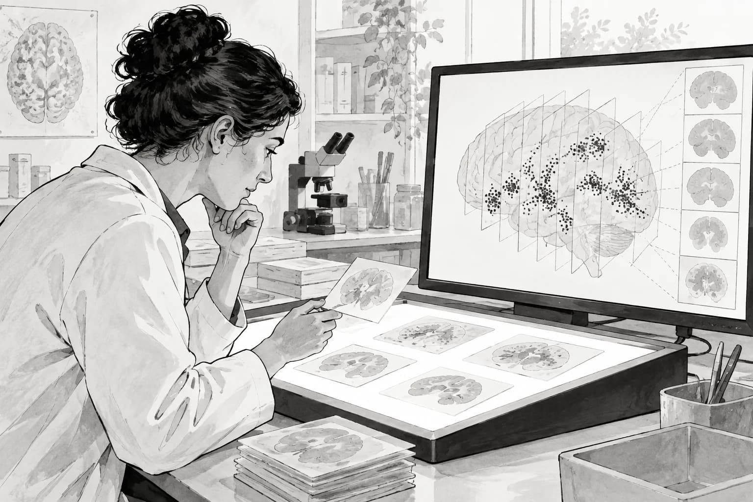
A closer map of damaged tissue
Neurofibrillary tangles are abnormal collections of tau protein found in brain tissue affected by Alzheimer’s disease. Researchers can study their number, location and shape on stained tissue slides, but drawing a precise boundary around every tangle takes a great deal of expert time. [1]
What the researchers tested
The training material came from post-mortem temporal-cortex slides from Alzheimer’s disease cases at three US institutions. The segmentation model was trained on 45 selected regions from 15 whole-slide images. Its held-out test set contained 664 tangles from 20 regions across seven slides. [1] [1]
Fine boundaries
The segmentation model estimates the outline of each tangle, which supports later measurements of shape and distribution.
Faster boxes
A separate object detector was about 60 percent faster, but it marked coarser boxes rather than pixel-level boundaries.
At whole-slide level, the automated scores were moderately correlated with separate expert ratings. That comparison supports the system as a research measurement tool. It does not show that the model can diagnose a person or predict how their symptoms will change. [1]
Where AI saves effort
The useful change is in the labeling bottleneck. An expert can mark the center of a tangle much faster than tracing its full edge. The pipeline converts those points into provisional masks, learns from them, and lets the expert concentrate on correcting likely misses. [1] [1]
Processing a full slide still required substantial computing. The paper reports about 32 minutes per slide for segmentation on one RTX 3090 GPU, plus conversion and post-processing. Faster than manual tracing does not mean instant or inexpensive in every laboratory. [1]
What remains uncertain
The data covered mature tau tangles in one brain region and used a specific stain. Other tau structures, brain regions, tissue preparation methods and scanners may behave differently. The authors expect the approach to transfer, but that still needs testing by other groups. [1]
The paper releases images, annotations, model weights and code, which makes reproduction possible. The next evidence to watch is whether independent laboratories can obtain similar measurements and whether these detailed maps reveal relationships that simpler pathology scores miss. [1]
Sources & context
One original study. The archive and publisher links refer to the same paper, not independent validation.
Learning precise segmentation of neurofibrillary tangles from rapid manual point annotations
Albarghouthi and colleagues · Scientific Reports · August 28, 2026